
Vexev: Innovation at the Intersection of Vascular Disease, Robotics, Imaging, and Personalized Diagnostics
The founders of Vexev sat down with Neeraj Hablani, for an interview exploring their vision to shift the approach of vascular medicine from reactive to proactive. That starts with better, simpler imaging.

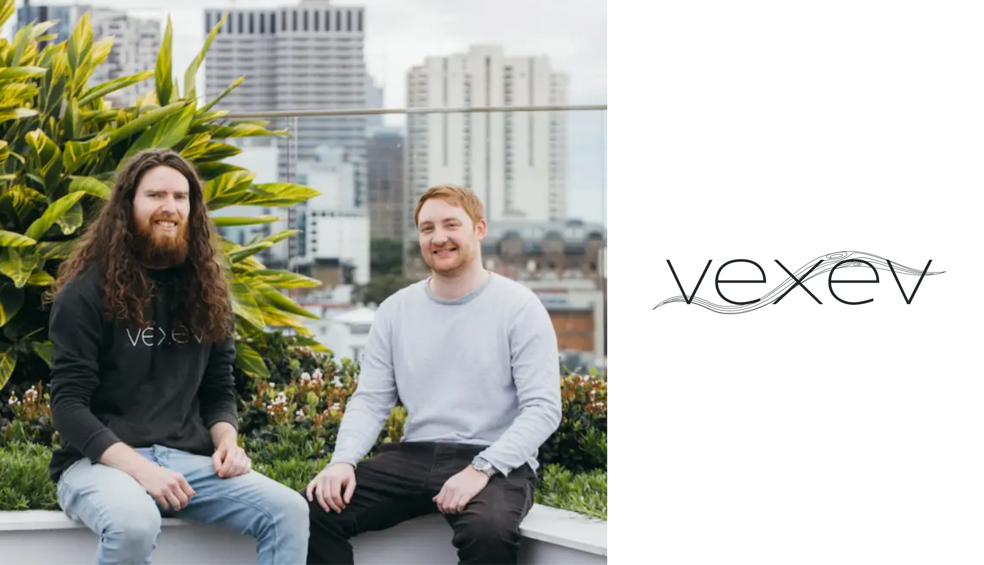
Dr. John Carroll and Dr. Eamonn Colley co-founders of Vexev sat down with Neeraj Hablani, Partner at Neotribe and a member of Vexev’s Board of Directors. Below is an excerpt from their discussion. Vexev’s vision is to shift the approach to vascular medicine from reactive to proactive. That starts with better, simpler imaging.
Vexev is hiring for a number of roles — you can find their open positions on their website! . If you are or know of a clinician or patient, their team would love to hear their story as it relates to vascular access management or peripheral artery disease
Introducing John and Eamonn
Neeraj Hablani: John and Eamonn – always a pleasure to chat. Thanks for taking the time to share your story and shine light on what the team at Vexev is up to. As a starting point, why don’t you two introduce yourself and share a bit about yourselves.
Dr. John Caroll: Thanks Neeraj! I grew up by the ocean in Sydney, Australia and loved math and physics at school. I always found the human body fascinating and thought it the most impressive ‘machine’ ever built. I was initially interested in pursuing medicine, but there wasn’t enough math, so I decided instead to pursue biomedical engineering at university.
Dr. Eamonn Colley: Great to chat Neeraj! I’m originally from Perth, Western Australia but have now lived in Sydney for the last 8 years. I had quite a lot of interests in high school, but one of my favorite subjects was mathematics. There was an interesting problem that the teacher gave us, which was to come up with a method for measuring the length of the coastline of Australia. It became apparent that the coastline length would increase as the measurement unit decreased (known as the coastline paradox), and so to describe this geometric shape we were introduced to a relatively new branch of mathematics, first published by Mandelbrot around the 1970’s, called fractal geometry. It was incredibly fascinating to learn more about fractals, especially how prevalent it is in nature but also the various applications in which the algorithms could be used in computation - a great example is in making the Pixar films! This love of understanding the world around us from a mathematical perspective and applying this knowledge led me to study Physics at university.
The Draw of Vascular Fluid Dynaics
NH: Mathematics and modeling are clearly a unifier for you two. You both met during your PhD program at UNSW nearly a decade ago. What attracted you both to graduate school to study vascular fluid dynamics?
JC: In my undergraduate studies I was mostly drawn to biomechanical modeling, that is using math to describe the behaviors of major systems within the body. Everything from simulating the heart as it beats, to leg movement dynamics. But what I loved the most was vascular flow modeling, as it combines fluid dynamics and structural physics while being an important part in understanding a critical component of our biology. I remember getting quite hooked on this area from the outset and haven’t since looked back!
I embarked on a PhD because I wanted to go further in understanding how the flow dynamics of blood and vascular disease are linked, as these links would have enormous potential in diagnostics and prevention. I also appreciated working with real patients as I was regularly reminded why solving this problem matters.
EC: During my Physics degree, I was fortunate to have the opportunity to study some really great subjects such as quantum mechanics, electromagnetism, fluid mechanics and solid-state physics. I really enjoyed them all, but I had to choose a research project in my Honors year, and I got drawn into the complexity of how to model the behavior of flow around objects.
My research used computational fluid dynamics to model a propeller rotating and the effects of cavitation, which is when water at a very low-pressure region changes state into a vapor bubble and then implodes when it travels to a higher pressure region. Very cool mathematics and had fun building the simulation, but the application was rather dry. I knew I wanted to continue in this area of computational fluid dynamics and so I applied for a PhD program at UNSW to research vascular fluid dynamics, which seemed like a very intriguing application and as John mentioned - an important part in understanding disease mechanisms and improving people’s lives.
Motivation to Start Vexev
NH: What you both highlight is a desire to translate mathematics and theory into something applied and useful for humanity – an inspiring unifier! What did you discover during your tenure at university that prompted you to create Vexev?
JC: Eamonn and I both started our PhDs on the same day and as you can probably tell by now, our interests were very aligned. I had done prior work in the vascular fluid dynamics area with my honors thesis, but this was using very basic approaches to modeling vessels. They were basically pipes. This approach was pretty standard, even across the current literature, but it was obvious that not simulating the real thing was going to be lacking in providing useful insights.
So for our research, we both agreed we wouldn’t take that direction and instead worked together to build a low-cost 3D ‘freehand’ ultrasound system, allowing us to safely scan patients’ vessels in 3D. We tracked patients over time with our 3D imaging system to scan their vessels and reconstruct models of the blood flow moving within.
EC: We actually built one of the largest data sets of its kind that captured actual cases of vessels remodeling and the onset of disease. Other researchers who did take the same direction in terms of using patient scans were limited by risks and costs associated with other imaging modalities (e.g. MRI or CT), meaning they couldn’t take regular weekly scans, or have a large enough set of patients. Both of these turned out to be crucial.
At the start of our study, we couldn’t know which patients would develop disease so we had to cast a wide net and have many patients scan regularly, which again was only possible with the system we had built. This also meant that we had the first dataset to show patients developing disease over time, instead of having disease already present in the first scan, which would obviously make it difficult to determine causation.
As we had the 3D blood flow behaviors for each of our own scans, we could use that to analyze what types of blood flow patterns led to the disease and eventually predict it. So this also gave us the opportunity to use our data to test and explore proposed theories in the literature, as well as devise some of our own.
We also found that it was important not only to take a snapshot of what the disease looks like at one point in time but that the true power was in tracking the rate of the progression of disease. Especially in the context of clinical decision-making.
This study, while not the original intention, effectively had us ‘monitoring’ patients, which was not normal practice. But here we saw cases that otherwise would have been missed, as symptoms only emerge at the late stage to prompt that first scan.
JC: Yes, we saw many times over patients having their first ‘scan’ after disease had already long set in, meaning they had fewer and worse options for intervention. And this ‘scan’ that they would get was extremely limited, mostly in the fact that it was 2D images, which was clearly in stark contrast with our 3D models and flow visualizations.
Altogether, as a result of this study and experience, we believed that the ‘monitoring’ could have a massive impact on many patients if we could just deliver these superior outputs at a greater scale.
This led us to create Vexev, so we could rebuild ultrasound imaging to make it more accessible to have a scan (e.g easy, safe and low cost) and to give better 3D outputs similar to CT/MRI (through automation and improved image processing), in order to solve this problem at scale. Hence the ‘better, simpler imaging’ being the first step in our mission.
The Status Quo of Vascular Imaging
NH: Makes sense – early detection is dependent on high-quality imaging. Let’s take a moment to dive into the biology and status quo of imaging. Our blood vessels help with the delivery of oxygen and nutrients while helping to eliminate waste from the body. Vascular disease is described as anything that impacts blood vessel functionality. Prior to exploring Vexev’s solution, can you walk us through the current standard of vascular imaging?
JC: Yeah great question. Many would likely be surprised that vascular imaging, which is mostly performed by an experienced ultrasound operator with a handheld probe, is very limited. The scanning process involves taking 2D image ‘snapshots’ with the ultrasound probe while the patient is present, which are then used to either fill in a template diagram or sometimes even hand-drawn diagrams to communicate the problem to vascular surgeons. Meaning that today, surgeons are going into theater and planning their interventions using these templates and hand-drawn diagrams.
As a result, patients who don't live near major hubs and who don't have access to an experienced sonographer are faced with much lower quality of care and the idea of imaging-based monitoring or surveillance becomes completely infeasible. This is the accessibility problem.
EC: There are some instances where patients use CT/MRI 3D imaging, which is obviously higher in quality but also much higher in cost, as well as requiring the injection of toxic contrasting agents to illuminate the vessels. Using CT/MRI is also completely infeasible for routine monitoring or surveillance, instead being suitable for diagnosis once a problem has already been found and the exposure risks to the patient are determined to be worth it.
Vexev’s Opportunity to Disrupt the Status Quo
NH: This is a good opportunity to segue into the Vexev mission statement. Talk us through your goals as a company and how Vexev plays a role in the detection and diagnosis of vascular disease.
JC: We started Vexev as we believed that accessibility and quality are the most important factors in using imaging as a preventative measure, rather than the end-stage diagnosis. To that end, we are setting a new standard for medical imaging in response to the barriers that prevent imaging today from being used for prevention.
Our device sets out to prove that having the imaging accessible to patients, allowing for regular scanning, means we will capture disease progression in its earlier stages. This gives surgeons far more options to intervene at a lower cost with greater chances of success. Further, as we scan more patients and build up a dataset, we’ll be introducing further metrics based on flow-related biomarkers that we uniquely capture.
We are starting with vascular disease as that is one of the largest growing problems for humans today, but as our device can be applied more generally for 3D medical imaging, we will apply this approach to all conditions which can be prevented through imaging-based early detection.
Innovating on 3D Ultrasound Imaging
NH: Your approach is a combination of hardware (HW) and software (SW) – and your first product, the Vexev Wave, is being manufactured as we speak. Which imaging modality does Vexev leverage? Walk us through the HW subcomponents.
JC: Our hard constraints for the device from the outset were that it needed to be simple enough for anyone to use as well as being safe enough and low cost (in terms of cost to scan) enough for unrestrained regular usage.
Ultrasound was selected as the modality due to safety and cost, but we are also able to generate outputs that are far more insightful than standard 2D ultrasound images. For example, we can output 3D interactive vascular geometries (which normally require CT/MRI imaging) and 4D flow reconstruction to visualize the flow over each heartbeat cycle. This gives clinicians and especially vascular surgeons plenty more information to work with.
This is achieved through our unique setup, where we use an ultrasound-guided robot to automate the scanning procedure, which also provides positional data allowing us to generate MRI/CT-like 3D images. Additionally, we are programming our ultrasound to take far more data samples and carry out major processing operations offline, rather than restricted to real-time which most ultrasound devices are today. Realtime makes sense if a human is guiding the probe, but not for a robot.
Leveraging Software and Data
NH: Equally important is your SW layer to the business. As part of your approach, Vexev will use ML to predict the health of arteries and veins. Walk us through your SW approach and how this further distinguishes the Vexev approach.
EC: Yes, very important as any ML application is only as good as the data you give it. While we will launch with a base set of features that are more focussed on detection and tracking as a form of monitoring, we will also be accumulating high quality data to learn what imaging and flow-based features can be used to predict vascular disease. So rather than just detecting disease, we will utilize our dataset and ML to determine what is likely to happen next and what can be done to avoid these scenarios.
The idea here is to understand on a patient level what the best treatment should be, and build a platform that clinicians can use to treat patients better, increase longevity for patients and for researchers to advance the field of medicine.
Apart from vascular health, we imagine many further applications, from intelligent triaging and patient management to surgical planning and eventually vascular optimisation.
Starting with a Problem Affecting 7% of the US Population
NH: Your peripheral arteries -- the blood vessels outside of your heart -- can build up plaque and thus cause less blood to flow to your tissues. This is known as peripheral arterial disease and is one of the focus areas for Vexev. Talk us through why you focused on this condition and how widespread of a problem peripheral arterial disease is.
JC: Yes, peripheral artery disease (PAD) is extremely widespread, with the prevalence of PAD estimated to be 7% of the US population (i.e >8.5 million adults), which is genuinely frightening. PAD in most cases refers to the vessels in the legs, where blood must travel the furthest path to these extremities. As these vessels taper with length, the smaller end vessels are more susceptible to narrowings and blockages.
EC: Adding to John’s figures, due to the fact that PAD doesn’t present with symptoms until it becomes advanced enough to start restricting blood flow, it is believed to be underdiagnosed by 40% - which is even scarier.
The underlying cause of PAD is atherosclerosis, which is the buildup of fatty plaques on the vessel wall creating a narrowing. This occurs all throughout the vascular system and is also the cause behind heart attacks. So if there is a buildup in the coronary arteries that leads to a narrowing, this creates a very high risk for a small piece of plaque or thrombus (i.e clot) to create a blockage. When this blockage happens, that is a heart attack.
So while PAD is a major issue in itself in terms of prevalence, this is an example of where we can also use the data from this application and apply learnings to other conditions such as heart attacks and strokes. We will have the leg scanning configuration of the Vexev Wave placed in point-of-care sites so that PAD detections can be made at an earlier stage, giving the patient and their clinician better options to prevent morbidities associated with late-stage PAD.
Making a Difference for Dialysis Patients
NH: Reducing that undiagnosed metric is a key opportunity for novel imaging techniques like Vexev. Expanding the conditions Vexev can diagnose is also a key opportunity; in addition to peripheral artery disease focused on the legs and extremities, walk us through how the arm scanner will have an impact.
JC: Yes correct, PAD is mostly relevant for the leg. Our arm scanning is focused more on the >500,000 hemodialysis patients in the US who receive dialysis through the vessels in their arm.
For patients on dialysis, the gold standard is to use a surgically created arteriovenous fistula (AVF). An AVF is created by joining a vein to an artery in the lower or upper arm to effectively create a short circuit to facilitate a high volume of blood flow for efficient dialysis.
There are a number of steps to having an AVF created and ready for dialysis, involving mapping the vessels with imaging, followed by the surgical creation procedure then followed by a 6-8 week ‘maturation’ process where the vein remodels to become artery-like and support a blood flow rate of 600 mL/min to enable efficient dialysis.
Unfortunately, probability states that 1 in 2 AVFs will require additional surgical procedures within 18 months of creation.
By having our arm scanning Vexev Wave configuration in the dialysis clinic, we can facilitate all the steps to getting patients onto a well-functioning AVF, as well as maintaining the AVF. The difference in cost between an intervention from an early detection vs late stage detection can be in the tens of thousands, but most important is the higher chance of success with an early intervention sparing further pain for the patient.
Impact to patients
NH: The sheer number of emergent procedures required after AVF creation is astounding and also an opportunity for Vexev to have an impact on hemodialysis patients globally. How do you hope high-quality imaging will impact patients’ lives with Vexev’s technology?
EC: Vexev is looking to normalize using high-quality medical imaging for routine monitoring as a form of late-stage disease prevention. For patients, this means that they will have regular and up-to-date information as to their current state, as well as procedures becoming more pre-emptive, less invasive, and having higher rates of success. This should give patients greater peace of mind and ultimately quality of life, as they avoid the morbidity associated with advanced vascular disease.
This also extends further from just vascular applications. Our vision is that Vexev’s technology will grow from vascular management to become a more holistic health management tool aided through maintaining our accessible high-quality imaging, as we expect to cover many further chronic health conditions.
We expect that Vexev will play a role in people’s lives well before they become seriously ill, as we expect the future of medicine to favor prevention once these tools are available and proven.
NH: Hence the mission of shifting vascular medicine from reactive to proactive. Thank you both so much for your time and for sharing your inspiring work, John and Eamonn.
EA: Thank you, Neeraj, it was a pleasure speaking with you.
JC: Thanks, Neeraj.